
When we think about Alzheimer’s disease, amyloid plaques and tau proteins usually come to mind. But they are not the whole story. Deep inside the brain, microglia are constantly at work. These immune cells help clear damaged material, respond to problems and keep the brain’s environment in balance. In Alzheimer’s disease, that routine starts to change. Microglia can remain switched on for too long, while their ability to clear unwanted material may decline. This shift has brought microglia, inflammation and the brain’s immune response into sharper focus.
A 2026 study has now added PTP1B to the conversation. Scientists found that this protein may act as a brake on certain microglial functions, with links to SYK signaling and amyloid-beta clearance in mouse models. The findings are early, but they raise an interesting question: could helping microglia work more effectively become part of future Alzheimer’s treatment? To put that question in context, we’ll look at how microglia work, what changes in Alzheimer’s, and where PTP1B research stands today.
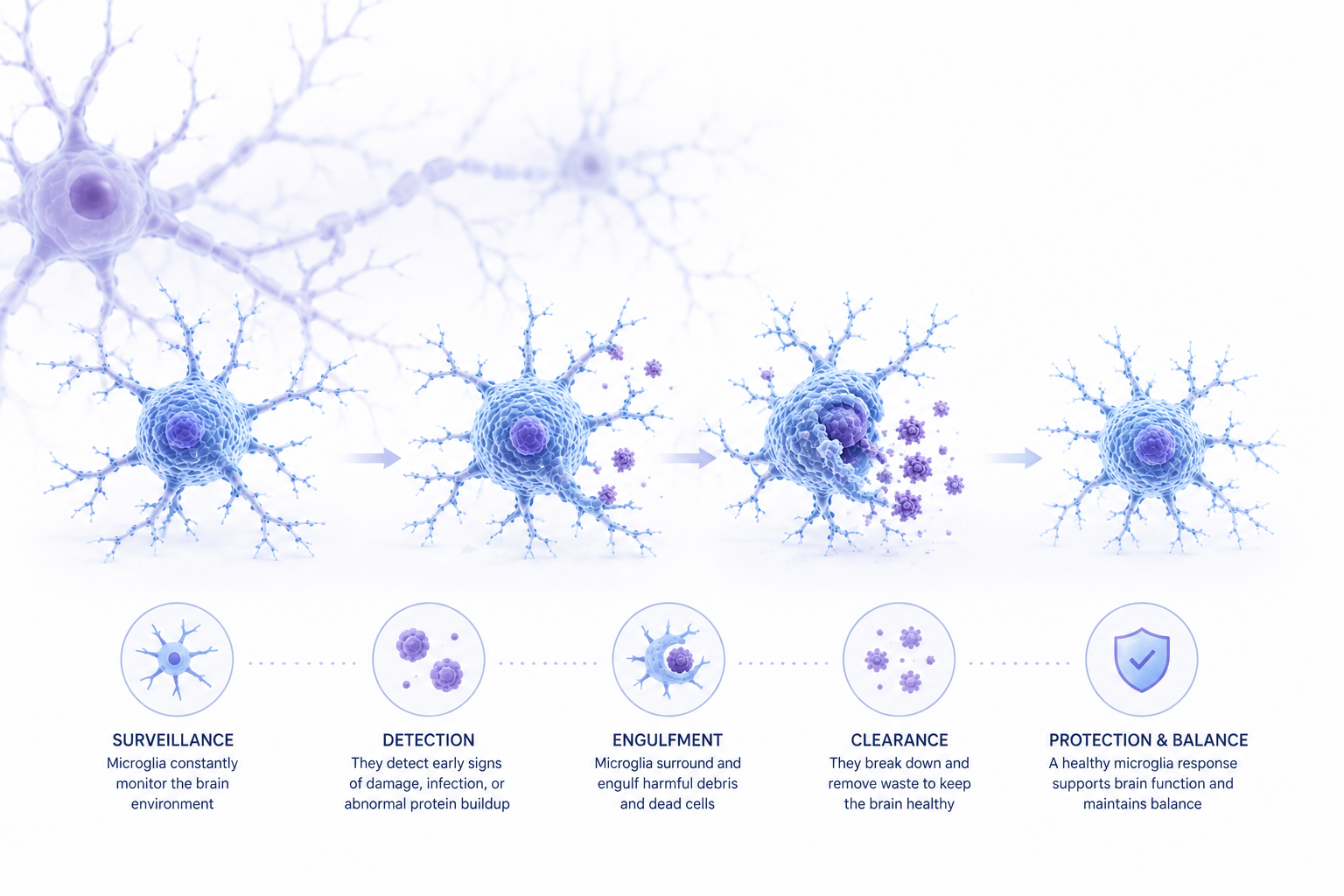
So, are microglia immune cells? Yes. Microglia are specialized immune cells that reside within the central nervous system and form an important part of the brain’s innate immune system. They continuously monitor their environment and respond when they detect damaged cells, foreign material, or other signs of trouble.
A useful way to picture microglia cells is as a cleaning and maintenance crew working throughout the brain.
In a healthy microglia brain environment, these cells perform several important functions:
This cleanup role becomes particularly relevant when researchers look at amyloid beta, a protein that can accumulate and form plaques associated with Alzheimer’s disease. Microglia can recognize and help clear Aβ material under normal conditions.
The problem is that the cleaning crew does not always remain effective when the brain is exposed to persistent disease-related stress.
Research increasingly points to a complicated relationship between microglia Alzheimer's disease pathology and inflammation.
When microglia encounter amyloid deposits, they become activated and move toward areas of damage. Initially, this response can be protective. However, prolonged activation can alter microglial behavior. Instead of efficiently clearing debris, the cells may become less effective at phagocytosis, the process through which they engulf and remove unwanted material.
This is one reason microglia inflammation has become an important area of Alzheimer’s research
Persistent activation can also contribute to the release of inflammatory signaling molecules, helping sustain neuroinflammation. In the Alzheimer’s brain, this creates a difficult cycle: disease-related proteins stimulate immune activity, while prolonged immune activation may contribute to further damage.
The relationship with amyloid and tau is also important. When clearance mechanisms become less effective, deposits of beta amyloid can accumulate. Microglial dysfunction may also influence the environment in which tau pathology and neurodegeneration progress. NIH research describes impaired microglial clearance and chronic neuroinflammation as important areas of investigation in Alzheimer’s disease.
In simple terms, the cleaning crew is still present—but it may no longer be picking up the waste efficiently.
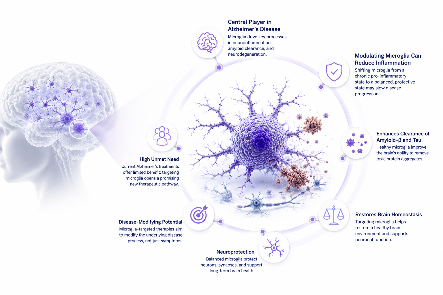
This change in thinking has encouraged researchers to ask a different question: rather than simply adding a new treatment, can existing microglial functions be restored or redirected?
Several signaling pathways are being investigated.
Triggering receptor expressed on myeloid cells 2 (TREM2) is a receptor involved in microglial function. It helps microglia respond to signals associated with damage and supports their response to amyloid-related pathology. Genetic changes affecting TREM2 are also associated with Alzheimer’s risk.
Think of TREM2 as a sensor telling the cleaning crew where attention is needed.
CSF1R signaling helps regulate microglial survival, maintenance, and activity. Because it influences the population and behavior of microglia, it has become another pathway of interest in research into neurodegenerative disease.
It can be thought of as part of the system deciding how many members of the crew are on duty and how they function.
Spleen tyrosine kinase (SYK) is another signaling protein involved in microglial responses, including phagocytosis. Researchers are investigating how controlling this signaling could influence the ability of microglia to respond to Alzheimer’s-related material.
Other pathways, including CD33, P2Y12, and complement-related signaling, are also being studied for their roles in microglial biology and Alzheimer’s disease.
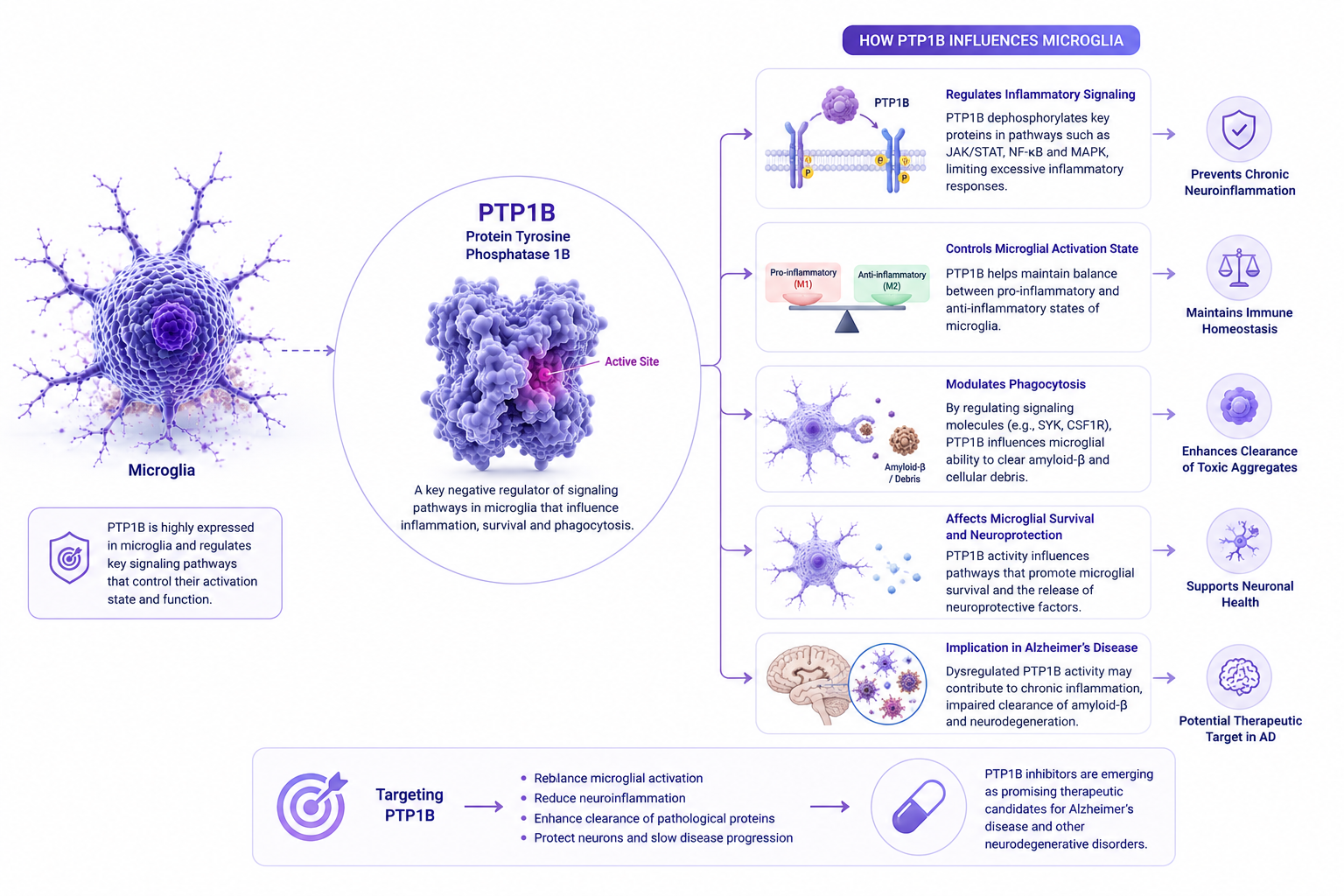
One of the more recent developments involves Protein Tyrosine Phosphatase 1B, commonly known as PTP1B.
PTP1B is an enzyme belonging to the protein tyrosine phosphatase family. These enzymes regulate cellular signaling by modifying proteins involved in communication inside cells. In the new Alzheimer’s research, PTP1B appears to act as a regulatory brake on microglial activity.
A 2026 study by Yuxin Cen and colleagues, published in Proceedings of the National Academy of Sciences, investigated PTP1B in Alzheimer’s disease models. The researchers found that genetic deletion or pharmacological inhibition of PTP1B improved memory-related outcomes and reduced amyloid-beta burden in APP/PS1 mice.
The study also identified an important connection between PTP1B and SYK signaling. The researchers reported that SYK is a direct substrate of PTP1B and that removing or inhibiting PTP1B enhanced signaling associated with microglial activation and phagocytosis.
That gives researchers a potentially interesting model: instead of forcing microglia into action, inhibiting PTP1B could help remove a molecular brake that is limiting their response.
The findings are particularly interesting because PTP1B has long been studied in metabolic disease, including pathways associated with insulin signaling. Alzheimer’s disease, obesity, and type 2 diabetes also share several metabolic and biological risk pathways, making the intersection worthy of further investigation.
However, the distinction between promising research and an available treatment is important. The PTP1B findings were demonstrated in experimental models, including APP/PS1 mice. They do not establish that a PTP1B-targeted treatment is effective or safe in people with Alzheimer’s disease. Human clinical trials would be required to determine whether the approach translates to patients.
PTP1B is only one part of a much broader research landscape. Current and emerging approaches include:
The FDA has approved amyloid-targeting therapies for appropriate patients with Alzheimer’s disease, demonstrating that disease-modifying approaches are moving from research into clinical practice. However, each treatment has specific eligibility requirements, risks, and monitoring considerations.
This section is provided for general educational purposes and does not represent Alzevita’s clinical claims or endorsement of any therapy. Treatment decisions should be made with qualified healthcare professionals.
As Alzheimer’s research becomes more focused on mechanisms, biomarkers and imaging can help researchers understand how disease changes over time.
MRI cannot directly show microglial activity. However, it can reveal structural changes associated with neurodegenerative processes. For example, hippocampal atrophy is associated with Alzheimer’s disease, and MRI-based measurements of hippocampal volume have been investigated as markers of disease-related change.
This makes quantitative imaging particularly valuable for research and monitoring. Repeated MRI scans can help assess changes in brain structure over time, while imaging and other biomarkers can support clinical research into disease progression and treatment response.
AI-assisted quantitative MRI could add another layer by helping analyze structural patterns consistently across scans. For tools such as Alzevita, the appropriate role is decision support—helping quantify and track relevant brain changes rather than making claims to diagnose or treat Alzheimer’s disease.
Alzheimer’s research is moving toward precision medicine, where treatment strategies are matched more closely to the biological characteristics of individual patients.
That could mean combining approaches that address amyloid, tau, inflammation, metabolism, and other disease pathways. It could also mean identifying biological changes earlier, before extensive neuronal damage has occurred.
Biomarkers may become increasingly important in this process. Imaging, blood-based biomarkers, cerebrospinal fluid measurements, and other tools are being investigated to identify disease-related changes and monitor progression.
The convergence of AI brain imaging and therapeutics could further strengthen this approach by helping researchers and clinicians interpret complex biological information.
The story of microglia Alzheimer's research is ultimately about balance.
Microglia are already equipped to patrol the brain, respond to threats, and clear unwanted material. In Alzheimer’s disease, that system can become disrupted. Understanding why it happens—and whether the process can be redirected—is now an important area of therapeutic research
The discovery of PTP1B’s relationship with SYK signaling adds an intriguing piece to that puzzle. In mice, removing this molecular brake improved microglial phagocytosis and reduced amyloid burden. But the findings remain preclinical, and much more research is needed before their relevance to human treatment can be established.
The goal may not always be to introduce something entirely new. Sometimes, it may be about understanding how to help the brain’s existing systems work more effectively.
PTP1B, or Protein Tyrosine Phosphatase 1B, is an enzyme involved in cellular signaling. A 2026 study found that inhibiting PTP1B enhanced microglial phagocytosis through SYK-related signaling in Alzheimer’s disease mouse models.