
At Alzevita, our mission is to simplify and standardise brain MRI segmentation and volumetric analysis through advanced, AI-powered image processing. We support radiologists, imaging labs, and researchers in generating fast, consistent, and reproducible brain volume data, streamlining workflows and enhancing decision support.
We envision a future where AI-driven medical imaging tools assist every radiology team in producing reliable, quantitative data, eliminating subjectivity and inefficiencies in traditional image analysis.
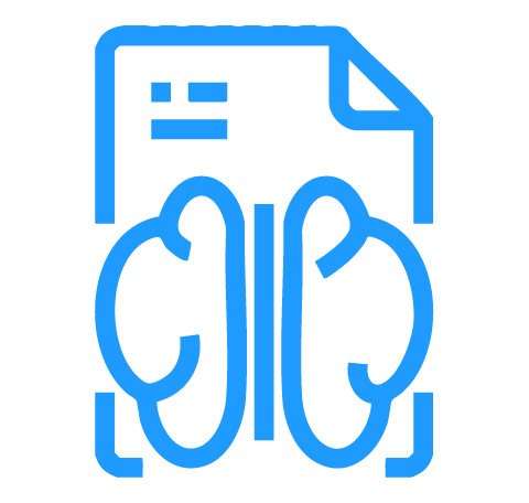
Alzevita is a cloud-based, AI-powered medical image processing software as a medical device intended to assist neurologists, radiologists and researchers with expertise in the analysis of 3D brain MRI scans.
Our system provides:
Built to reduce the need for manual segmentation, Alzevita helps neurologists, radiologists, and researchers deliver efficient, standardized, and reproducible outputs while remaining aligned with clinical and regulatory standards.

 Accuracy
Through
AI
Accuracy
Through
AI
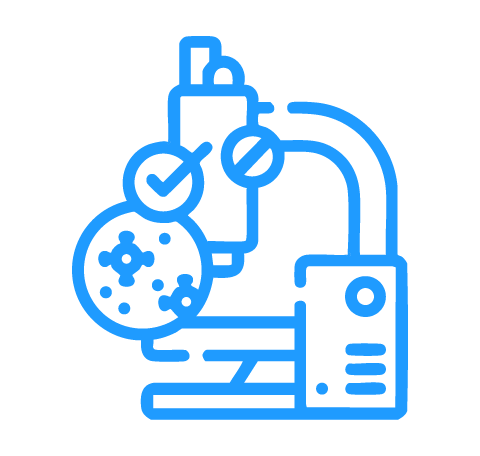
Delivering precise segmentation using validated AI models
 Efficiency &
Speed
Efficiency &
SpeedMinimising manual effort to optimise imaging workflows
 Reproducibility
& Standardisation
Reproducibility
& StandardisationEnsuring consistent volumetric outputs across cases and timepoints
 Compliance First
Built for professional use
Neuroradiologists seeking quantitative insights to enhance interpretation and reporting

Automating brain MRI processing for radiologists

Researchers focused on neuroimaging and structural brain analysis
Hospitals and MRI centers integrating AI into radiology workflows for standardized analysis
Everything you need to know about Alzevita
Our Commitment
We are committed to enabling accurate, scalable, and reproducible brain imaging through trusted, clinically focused software. Alzevita is designed to assist neurologists, radiologists and researchers with consistent quantitative outputs, without making diagnostic claims or replacing clinical judgment.